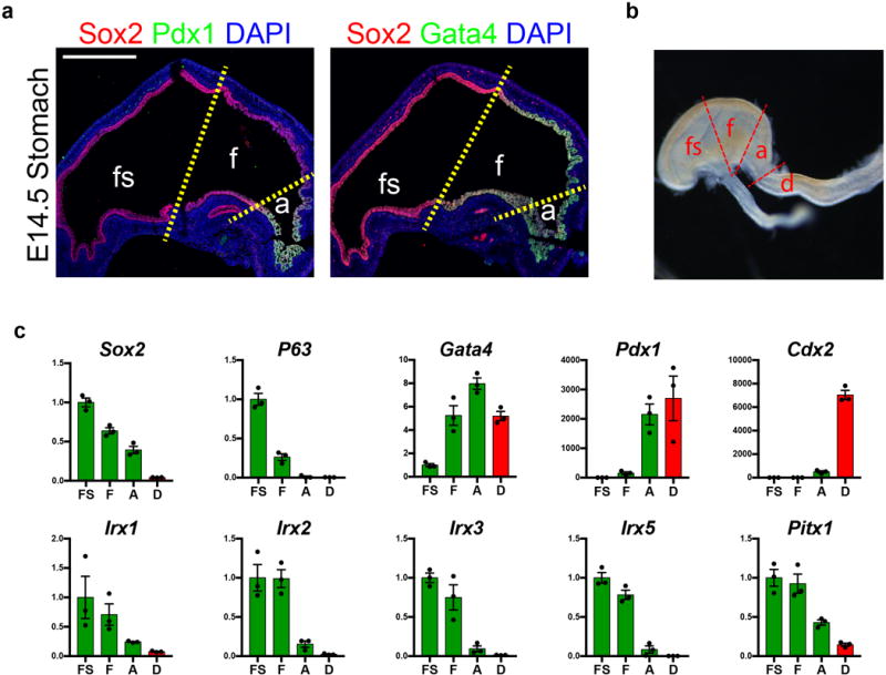
Extended Data Figure 1. Defining molecular domains in the developing stomach in vivo.

a, Analysis of Sox2, Pdx1, and Gata4 in the embryonic mouse stomach (E14.5) showed that the fundus (f) was Sox2+Gata4+Pdx1−, whereas the antrum (a) was Sox2+Gata4+Pdx1+. The forestomach (fs) expressed Sox2 but neither Gata4 nor Pdx1. b, Brightfield stereomicrograph showing dissected regions of the E14.5 mouse stomach that were analyzed by qPCR. fs, forestomach; f, fundus; a, antrum; d, duodenum. c, Dissected regions in b were analyzed by qPCR for known regionally expressed markers (Sox2, P63, Gata4, Pdx1, and Cdx2) to validate the accuracy of micro-dissection. qPCR analysis of the dissected E14.5 stomach regions showed that putative fundus markers Irx1, Irx2, Irx3, Irx5, and Pitx1 were enriched in the fundus compared to the antrum. n=4 biological replicates per dissected region. Scale bar, 500 μm. Error bars represent s.d.
