
Extended Data Figure 5. hFGOs contain organized glands supported by associated mesenchymal layer.
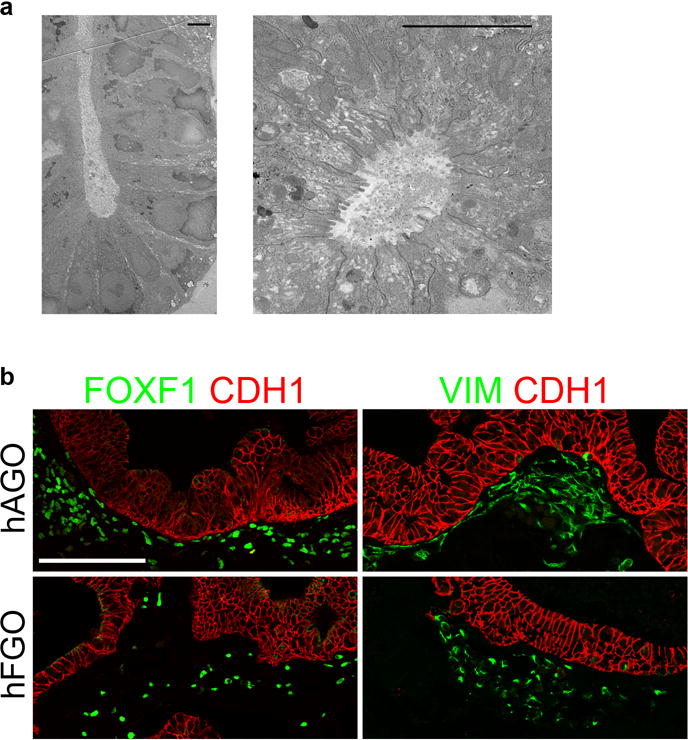
a, Transmission electron micrographs demonstrated that hFGO glands exhibited organized architecture with narrow apical membranes. b, Both hFGOs and hAGOs contained a supporting layer FOXF1+/VIM+ undifferentiated fibroblasts. Scale bars, 5 μm (a) and 100 μm (b).