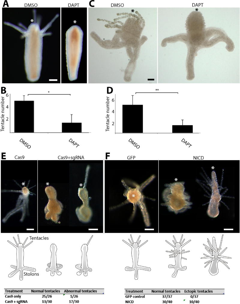
Figure 4. Notch signaling is required for tentacle formation.
(A, B) DAPT treatment following decapitation inhibits tentacle but not hypostome regeneration. (C, D) DAPT treatment during metamorphosis blocks tentacle development but has no effect on hypostome, body column or stolon formation. (E) CRISPR-Cas9 mediated mutagenesis results in defective tentacle patterning during metamorphosis. (F) Ectopic expression of NICD leads to the development of ectopic tentacles. Scale bars: 250 µm in (A), 40 µm in (C) 100 µm in (E) and (F). Asterisk marks the oral pole in all images. * Student t-test with p<0.0001. ** Student t-test with p<0.000001.

