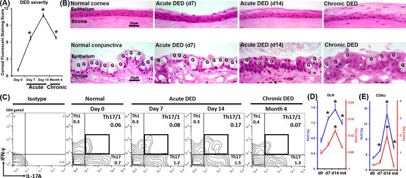
Figure 1. Increased IFN-γ+Th17 cells (Th17/1) in severe dry eye disease (DED).

Frequencies of Th1, Th17, and Th17/1 cells were examined by flow cytometry throughout the course of DED (normal, acute, and chronic stages). (A) DED severity was evaluated by corneal fluorescein staining scores at different stages (n = 20 eyes per time point). *, p < 0.05 as compared to Day 0. (B) Pathological changes of corneal (upper panel) and conjunctival epithelium (lower panel) in different stages of DED. Ocular surface in DED is characterized by change in epithelial thickness in the cornea, and loss of goblet cells (marked as G) in the conjunctiva. (C) Representative flow cytometry plots show cell frequencies gated on CD4+ cells. (D) Kinetic changes of Th17 (blue line) and Th17/1 (red line) cells in eye-draining lymph nodes (DLN) during the course of disease are summarized as mean±SEM from one representative experiment out of three performed (n = 4 mice per time point per group). (E) Kinetic changes of the Th17 (blue line) and Th17/1 (red line) cells in conjunctivae (CONJ) are summarized as mean±SEM from all three experiments with 4–6 eye tissues pooled together as one flow cytometry sample (n = 4 samples per time point per group). *, p < 0.05 as compared to Day 0.
