
Figure 2. Both Th17 and Th17/1, but not Th1 cells isolated from DED are pathogenic.
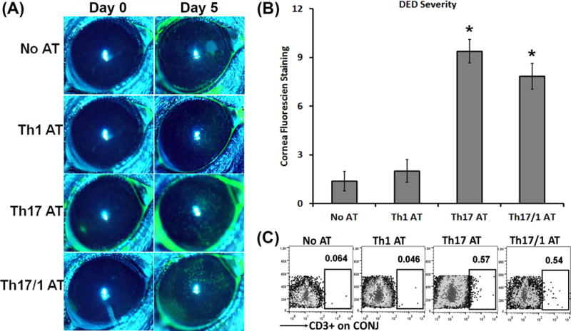
Severe DED was induced for 14 days in wild-type mice, and then Th1, Th17, and Th17/1 cells were isolated from draining lymph nodes. 1 × 10⁴ cells from each subset were intravenously injected into Rag1 KO mice. Immediately after the adoptive transfer, these Rag1 KO mice were subject to desiccating stress for 5 days. AT, adoptive transfer. (A) Clinical disease severity was evaluated by corneal fluorescein staining and representative images show baseline (Day 0) and 5 days post-AT. (B) DED scores in each group at Day 5 are summarized as mean±SEM in bar graphs (n = 6–8 eyes per group). *, p < 0.05 as compared to No AT or Th1 AT group. (C) Representative flow cytometry dot plots from three separate experiments (4–6 eye tissues pooled together for each group) show increased infiltration of transferred T cells in the conjunctivae (CONJ) in mice with AT of Th17 and Th17/1 cells.