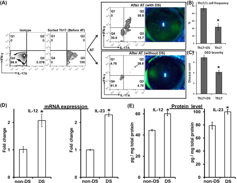
Figure 3. Th17 cells isolated from DED convert into IFN-γ-secreting cells associated with DED-induced IL-12 and IL-23.

DED-specific Th17 cells were isolated from severe DED (Before AT) and then adoptively transferred into Rag1 KO mice which were then subject to desiccating stress (DS) or not (non-DS) for 5 days. AT, adoptive transfer. (A) Clinical disease severity was evaluated by corneal fluorescein staining and representative images were shown 5 days post-AT. In addition, eye-draining lymph nodes of the recipient mice were collected and analyzed for IFN-γ and IL-17A expressions by flow cytometry (After AT). The data were summarized as mean±SEM for Th17/1 frequency (n = 3 mice) (B) and disease score (n = 6 eyes) (C) from one experiment out of two performed. *, p < 0.05. (D) mRNA and (E) protein expressions of IL-12 and IL-23 in the draining lymph nodes were quantified by real-time RT-PCR and ELISA, respectively. *, p < 0.05. Six eyes from 3 mice in each group were analyzed.
