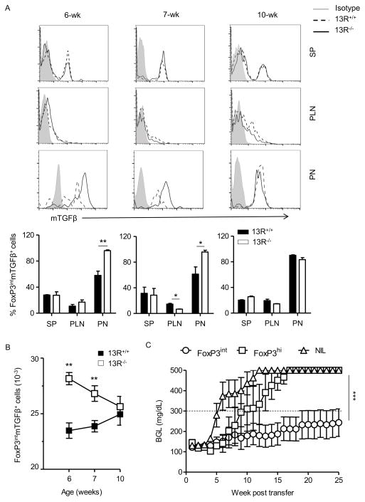
Figure 4. The frequency of protective FoxP3int Tregs is higher in 13R−/− than 13R+/+ mice.
Cells from the spleen (SP), pancreas (PN) and pancreatic lymph nodes (PLN) of 13R+/+FoxP3-GFP or 13R−/−FoxP3-GFP female NOD mice were stained with antibodies to CD4, CD25, and membrane bound TGFβ (mTGFβ) and the frequency of CD4+CD25+GFPint(FoxP3int)mTGFβ+ cells was analyzed by flow cytometry at the age of 6, 7 and 10 weeks. In (A) the histograms show representative FACS plots for expression of mTGFβ by cells gated on CD4 and CD25 and FoxP3int at the indicated weeks for the SP, PLN and PN. Each bar graph show the percentage ± SD of FoxP3int and mTGFβ expression by cells gated on CD4 and CD25 compiled from 3 independent experiments. (B) Shows the absolute number of FoxP3intmTGFβ+ cells ± SEM compiled from 3 independent experiments. *p<0.05, **p<0.005 as determined by two-tailed Student’s t-test. (C) Shows the mean BGL ± SEM in 13R+/+ NOD mice (8 per group) given sorted FoxP3int or FoxP3hi Tregs (4 × 10⁵ cells/mouse) at the age of 12 weeks. A group of mice that did not receive Tregs (NIL) was included for control purposes. ***p<0.0001 as determined by Kruskal-Wallis test.

