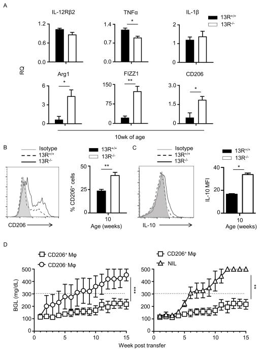
Figure 7. 13R−/− mice develop protective pancreatic macrophages which delay T1D upon transfer to 13R+/+ mice.
(A) CD11c−CD11b+ macrophages were sorted from the pancreas of 10 week-old female 13R+/+ and 13R−/− NOD mice after depletion of CD11c cells. RNA was then extracted by Trizol and used to measure mRNA expression for markers associated with M1 and M2 macrophages. Each bar represents the mean RQ ± SEM compiled from 3 independent experiments. *p<0.05, **p<0.005 as determined by two-tailed Student’s t-test. (B) Pancreatic cells from 10-week-old female 13R+/+ and 13R−/− NOD mice were stained with antibodies to CD11b, F4/80 and CD206 and the cells were analyzed for CD206 expression. The plots show a representative experiment for expression of CD206 by cells gated on CD11b+ and F4/80+. The bar graphs show the percent mean ± SD of CD11b+F4/80+CD206+ cells compiled from three independent experiments. **p<0.005 as determined by two-tailed Student’s t-test. (C) CD11b+cells were isolated from the pancreas of 10-week-old female 13R1+/+ and 13R1−/−NOD mice. The cells were then stimulated with LPS (0.5μg/ml) for 24 hours and stained with antibodies to CD11b, F4/80, CD206 and intracellular IL-10. The plot shows a representative experiment for IL-10 expression by cells gated on CD11b, F4/80, and CD206. The bar graph shows the percent mean ± SD of CD11b+F4/80+CD206+IL-10+ cells compiled from three independent experiments. *p<0.05 as determined by two-tailed Student’s t-test. (D) Pancreatic cells were harvested from 10 week-old 13R−/− female NOD mice and used to sort two populations of CD11b+F4/80+ macrophages (Mϕ) on the basis of CD206 expression. The CD206+ and CD206− Mϕ were separately transferred (5 × 10⁵ cells/mouse) into 12-week old 13R+/+ mice to test for resistance to T1D. The hosts given CD206+ (n = 9) and CD206− (n =8) cells were then monitored for BGL twice a week for 15 weeks. A group of mice (n = 7) which did not receive any cell transfer (NIL) was included for control purposes. A mouse is considered diabetic when the BGL is ≥300 mg/dl for three consecutive tests. **p<0.005 and ***p<0.0001 as determined by Mann-Whitney U test.

