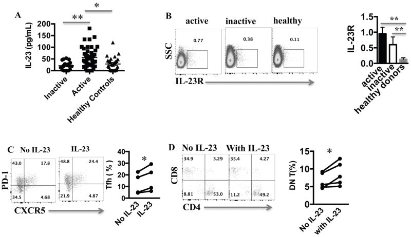
Figure 1. IL-23 is elevated in SLE and promotes the generation of Tfh and DN T cells.
(A) IL-23 level was measured from sera of healthy donors, active and inactive SLE patients. (B) CD3+ T cells from healthy donors, active and inactive SLE patients were stained with an anti-IL-23R antibody. A representative dot plot and cumulative results are shown here. (C) SLE T cells were stimulated with anti-CD3/CD28 antibodies with or without IL-23 for 5 days and then stained for the Tfh markers (CD3+CD4+PD-1+CXCR5+) (representative dot plot and cumulative results from 5 patients). (D) SLE T cells were stimulated with anti-CD3/aCD28 antibodies with or without IL-23 for 5 days and then stained with CD3, CD4 and CD8 (gated for CD3+, representative experiment and cumulative data from 5 patients). * p < 0.05, ** p < 0.01. Error bars represent SD.

