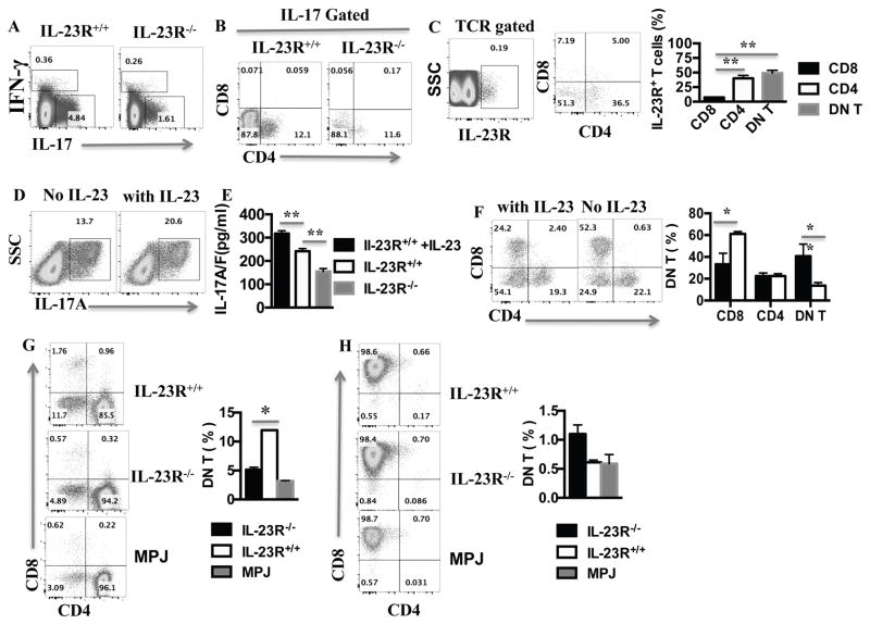
Figure 6. IL-23R deficiency limited the production of IL-17 and the generation of DN T cells.
Spleens were harvested from IL-23R−/− MRL.lpr and IL-23R+/+MRL.lpr mice as indicated, and single cell suspension was prepared. (A) IFN-γ and IL-17 produced by T cells were analyzed by intracellular staining and cytometry after gating on live TCRαβ+ T cells (see Experimental Procedures) (B) IL-17-producing T cells were analyzed for CD4 and CD8 expression by cytometry after gating on TCRβ+IL-17+; (C) TCRβ+ splenocytes from IL-23R+/+ MRL.lpr were stained with anti-IL-23R antibody and evaluated using flow cytometry (cumulative results (right panel), n=3 ). (D) Splenocytes from IL-23R+/+ MRL.lpr were stimulated with anti-CD3/anti-CD28 antibodies for 3 days with or without IL-23; then the cells were harvested to analyzed for IL-17 expression by cytometry via gating on TCRβ+ T; (E) The supernatants from the 3 day-cell cultures described in D including a IL-23R−/− control were used for IL-17A/F level measurement with ELISA (n=3 mice in each plot). (F) 4×10⁶ splenocytes from 8-week-old MRL.lpr mice were stimulated with anti-CD3/28 in the presence or absence of 25ng/ml IL-23 for 2 days; the cells were then harvested and analyzed for CD4 and CD8 expression by cytometry after gating on CD3+ T cells (representative plot of 3 mice-left panel and cumulative results-right panel). (G–H) CD4+ (G) and CD8+ (H) T cells were sort-purified from the spleens of 12-week-old IL-23R+/+ MRL.lpr, IL-23R−/−MRL.lpr and MRL.MPJ mice. They then were stimulated with anti-CD3/CD28 for 3 days before being (re)stained for CD3, CD4 and CD8 expression (representative experiment left panel, cumulative data from 3 mice/group, right panel). *= p< 0.05. Error bar represents SD. Data are representative of three independent experiments.

