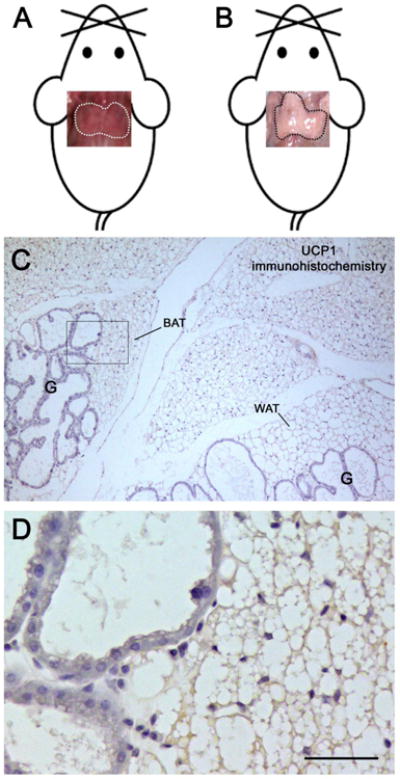
Figure 1.

The mouse anterior subcutaneous depot during lactation. Upper panels, pictures of the gross anatomy of the interscapular depot fitted on mouse templates. In non-pregnant female mice (A), the interscapular fat (dotted area) is easily recognized by its brownish color; after 10 days of lactation (B) it turns white and the depot acquires a white fat-like macroscopic appearance. In sections processed for immunohistochemistry (C), interscapular multilocular adipocytes containing large lipid droplets exhibit very weak UCP1 expression. Mammary gland alveoli (G) are found close to both brown (BAT) and white (WAT) adipose tissue lobules. At higher magnification (D), no clear boundaries are visible between the secretory epithelium and the adipose parenchyma; note the weak and homogenous UCP1 staining in multilocular adipocytes. D is the enlargement of the area framed in C.
Bar: 3 cm in A, 250 μm in B, 40 μm in C.
