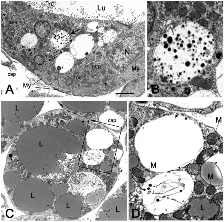
Figure 2.

Transmission electron microscopy images from specimens of peripheral anterior mammary gland in contact with interscapular BAT on day 1 of mammary gland involution. In A, the epithelial cells bordering the alveolar lumen (Lu) contain numerous mitochondria (m), stacked rough endoplasmic reticulum (RER), and large vacuoles containing milk protein secretory granules; the vacuole indicated by the arrow is enlarged in B. C: a multilocular adipocyte containing several “brown-like” mitochondria (M) packed with laminar cristae and two milk protein secretory vacuoles (enlarged in D). Cap: capillary, My: myoepithelial cells, N: nucleus, L: lipid droplet. Bar: 1 μm in A and C, 0,3 μm in B and D.
