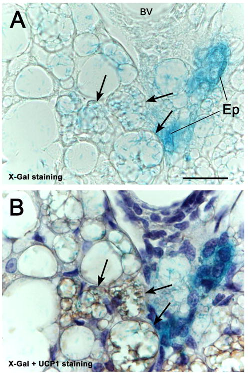
Figure 3.

X-Gal staining of the peripheral anterior mammary gland in contact with interscapular BAT of a WAP-Cre/R26R mouse 10 days post-lactation. In A, not only mammary epithelial structures (Ep), but also some multilocular adipocytes (arrows) show the blue X-Gal precipitate. UCP1 immunohistochemistry and hematoxylin counterstaining, performed in the same section (B), show that the X-Gal-positive multilocular adipocytes also exhibit UCP1 immunoreactivity (arrows). bV: blood vessel.
Bar: 25 μm.
