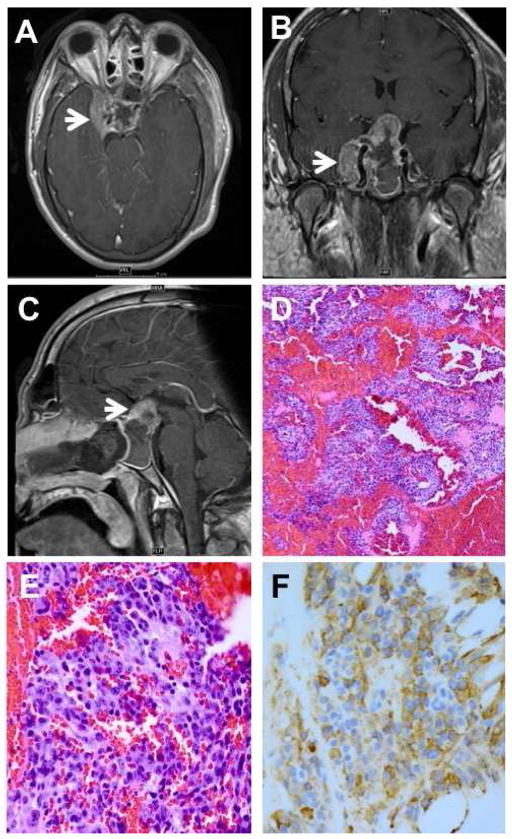
Figure 1.
A: MRI showing invasion of the right cavernous sinus. B: MRI showing encasement of the carotid artery (arrow) and compression of optic chiasm. C: MRI showing 4.1 cm sellar/suprasellar mass with sellar expansion. D: biopsy revealing pituitary adenoma with extensive hemorrhage. E: monomorphic population of amphophilic and sparsely granulated cells. F: adenoma cells show diffuse reactivity for human growth hormone. (D: hematoxylin and eosin, 25X; E: hematoxylin and eosin, 100X; F: anti-human growth hormone, 100X).

