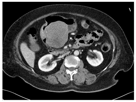
Figure 2.

Computed tomography scan image of a gastric gastrointestinal stromal tumors. A submucosal tumor measuring 7.7 cm × 7.6 cm × 7.2 cm in dimension was located on the posterior wall of gastric antrum. An ill-defined hypodensity within the mass could represent an area of necrosis.
