
Figure 1.
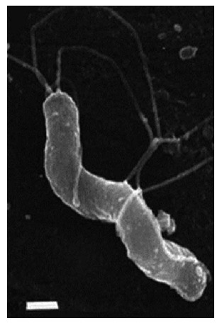
Morphology of Helicobacter pylori. S-shaped H. pylori with five to seven sheathed polar flagella. Field emission SEM, bar = 0.5 μm. (Field emission SEMs courtesy of L. Thompson and negative stains courtesy of S. Danon, School of Microbiology and Immunology, University of New South Wales). From: Helicobacter pylori: Physiology and Genetics. Mobley HLT, Mendz GL, Hazell SL, editors. Washington (DC): ASM Press; 2001. Chapter 6, Morphology and Ultrastructure[54].