Figure 3.

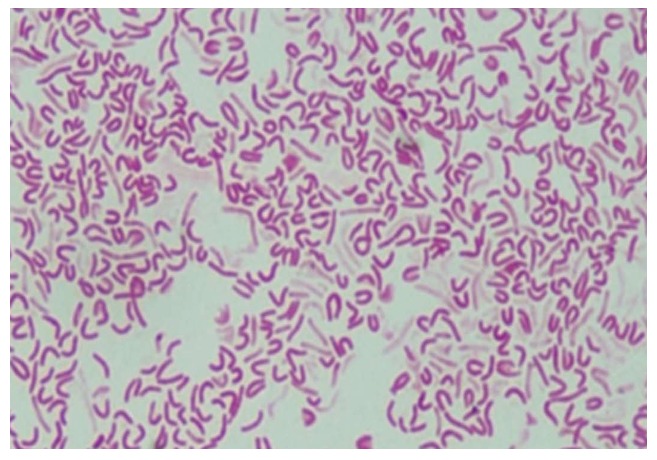
Microscopic images of morphological forms of Helicobacter pylori after exposure to antibiotics: S-shaped, U-shaped, C-shaped and coccoid-shaped. From: Faghri et al[65].

Microscopic images of morphological forms of Helicobacter pylori after exposure to antibiotics: S-shaped, U-shaped, C-shaped and coccoid-shaped. From: Faghri et al[65].