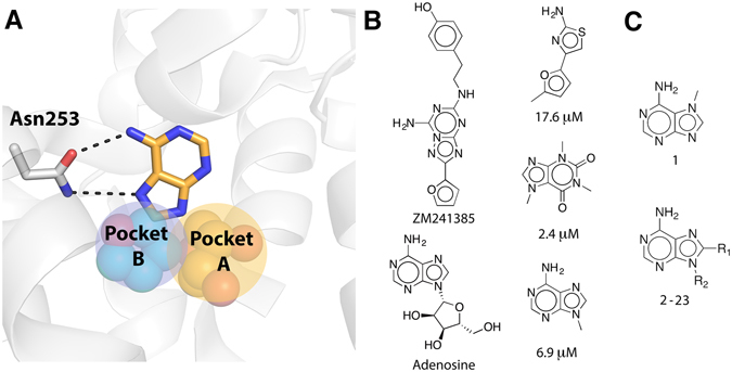
Figure 1.

(A) Orthosteric binding site of the A2AAR shown as white cartoon with Asn253 in sticks. The adenine group is shown in sticks with carbon atoms in gold and hydrogen bonds indicated with black dashed lines. Two adjacent subpockets are shown as spheres with yellow (pocket A, ribose group of endogenous agonist adenosine from the crystal structure with PDB code 2YDO)18 and cyan (pocket B, furan group of antagonist ZM241385 from the crystal structure with PDB code 4EIY)17 carbon atoms. (B) Two adenine-based and three fragment-sized ligands of the A2AAR. Ki values are provided for the fragment ligands21, 22, 24. (C) 2D structures of compounds 1–23. The R-groups are shown in Table 1.
