
Figure 1.
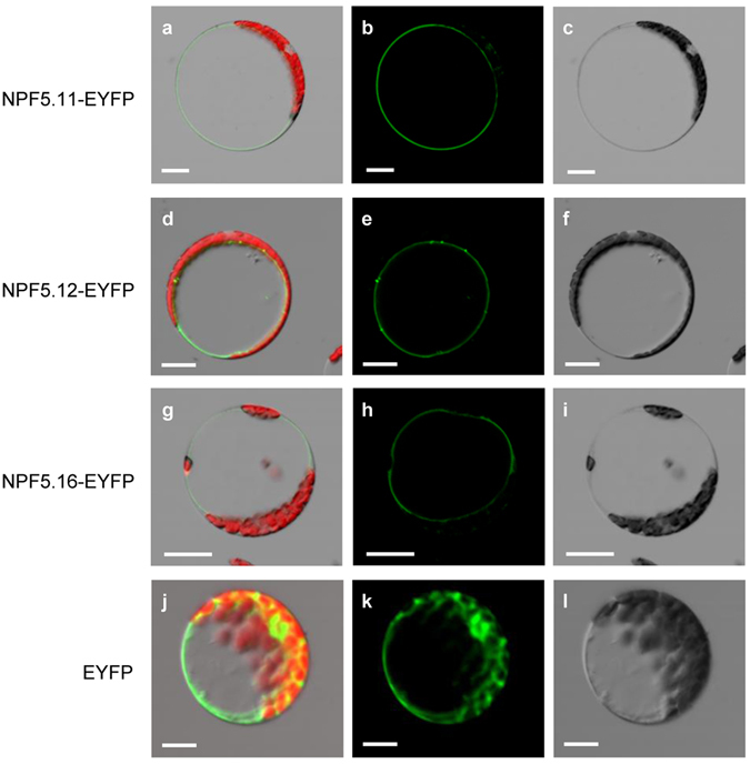
Subcellular localization of NPF5.11, NPF5.12 and NPF5.16. NPF5.11-EYFP (a–c), NPF5.12-EYFP (d–f), NPF5.16-EYFP (g–i) or EYFP (j–l) was driven by the cauliflower mosaic virus 35 S promoter and transiently expressed in Arabidopsis mesophyll protoplasts. Overlap images of EYFP (green) and chlorophyll (red) fluorescence (a,d,g,j), EYFP fluorescence (b,e,h,k), and bright-field (c,f,i,l) images are shown. Bars = 20 μm.