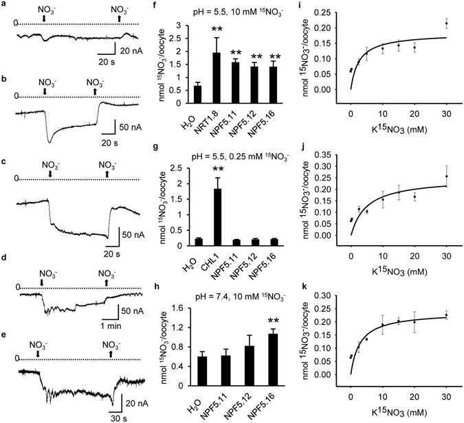
Figure 2.

Functional characterization of NPF5.11, NPF5.12 and NPF5.16 in oocytes. (a–e) Currents elicited in oocytes injected with H2O (a), CHL1 cRNA (b), NPF5.11 cRNA (c), NPF5.12 cRNA (d) or NPF5.16 cRNA (e). Oocytes were voltage clamped at −60 mV and representative inward currents elicited by 10 mM NO3 − at pH 5.5 were recorded. (f–h) Nitrate uptake activity in oocytes injected with H2O, NPF5.11 cRNA, NPF5.12 cRNA, NPF5.16 cRNA, NRT1.8 cRNA or CHL1 cRNA. Oocytes were incubated with 10 mM 15NO3 − at pH 5.5 (f), 0.25 mM 15NO3 − at pH 5.5 (g) or 10 mM 15NO3 − at pH 7.4 (h) for 12 h. Values are means ± SD (n = 8–12). Asterisks indicate difference at P < 0.01 (**) compared with the H2O-injected oocytes by Student’s t-test. (i–k) Uptake kinetics of NPF5.11 (i), NPF5.12 (j) and NPF5.16 (k). Oocytes injected with NPF5.11 cRNA (i), NPF5.12 cRNA (j) or NPF5.16 cRNA (k) were incubated with indicated concentrations of 15NO3 − at pH 5.5 for 1.5 h, and the 15N contents were determined. Values are means ± SD (n = 6–12). The Km was calculated by fitting to the Michaelis-Menten equation using a nonlinear least squares method in the SigmaPlot program. The Km was 2.57 mM, 4.84 mM, or 2.91 mM for NPF5.11, NPF5.12 or NPF5.16, respectively.
