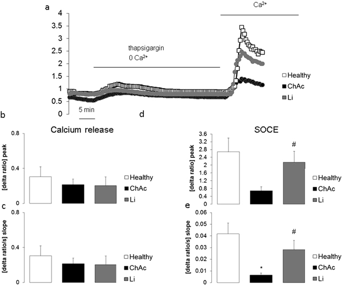
Figure 4.

Intracellular Ca2+ release and store-operated Ca2+ entry (SOCE) in neurons from healthy volunteers and from ChAc patients without or with lithium treatment. (a) Representative tracings of Fura-2 fluorescence-ratio in fluorescence spectrometry before and following extracellular Ca2+ removal and addition of thapsigargin (1 µM), as well as re-addition of extracellular Ca2+ in neurons generated from healthy volunteers (white squares) and from ChAc patients without (black circles) and with (grey circles) lithium (24 h, 2 mM) treatment. (b,c). Arithmetic means (±SEM, n = 37–74 cells from 4 individuals) of slope (b) and peak (c) increase of fura-2-fluorescence-ratio following addition of thapsigargin (1 µM) in control neurons (white bar) and in ChAc neurons without (black bar) and with (grey bar) lithium (24 h, 2 mM) treatment. (d,e) Arithmetic means (±SEM, n = 37–74 cells from 4 individuals) of slope (d) and peak (e) increase of fura-2-fluorescence-ratio following re-addition of extracellular Ca2+ in neurons from healthy volunteers (white bars) and in neurons from ChAc patients without (black bar) and with (grey bar) lithium (24 h, 2 mM) treatment. *(p < 0.05) indicates statistically significant difference to respective value in neurons from healthy volunteers, #(p < 0.05) indicates statistically significant difference to respective value in absence of lithium.
