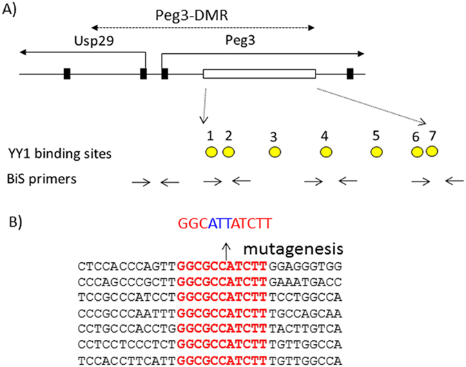
Figure 1.

7 YY1 binding sites localized within the Peg3-DMR. (A) The 4-kb genomic region corresponding to the Peg3-DMR is indicated with a dotted line with arrows. The 1st and 2nd exons of both Peg3 and Usp29 are indicated with thick vertical lines, whereas the 2.5-kb YY1 binding region is indicated with an open box. The relative positions of the 7 YY1 binding sites are indicated with circles, and the positions of several sets of BiS (Bisulfite Sequencing) primers for DNA methylation analyses are indicated with arrows. (B) The sequence of the 7 YY1 binding sites are shown along with the flanking sequences. The sequences of the 7 YY1 binding sites with red are identical to each other. The three bases of each YY1 binding site were mutated from 5′-GCC-3′ to 5′-ATT-3′.
