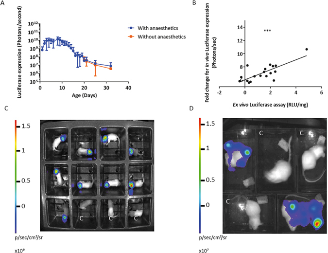
Figure 1.

Intracranial neonatal administration of VSV-G SFFV lentiviral biosensor, with mean bioluminescence quantified from anaesthetised and non-anaesthetised mice. Mice received the VSV-G pseudotyped lentivirus vector containing the constitutive SFFV promoter, at birth via intracranial injections (n = 9). They were imaged daily, without anaesthetic, until 21 days old. (A) Mean bioluminescence was quantified. The mice were then allocated randomly into two groups and whole-body bioluminescence imaging was undertaken either with or without isoflurane anaesthesia. There was no significant difference in luciferase expression between the two groups, (mean +/− SD, P = 0.657). (B) New born mice received an intracranial injection of VSV-G SFFV lentiviral vector. Over the course of development mice were bioimaged and brain tissue from three mice was collected at weekly time points for ex vivo luminometry (n = 3). A strong positive correlation was observed between the fold change of in vivo luciferase expression at the time of euthanasia and ex vivo luciferase expression, P = 0.0003, R2 = 0.5690. (C) Imaging of neonatal mice and (D) adult mice without anaesthetic, mice marked with C are control mice which have not received vector. The region of interest (ROI) used to quantify conscious neonatal mice in each enclosure, was 5 cm by 5 cm. Once the mice had reached adult hood the ROI for both conscious and unconscious mice was 9 cm by 12.5 cm. The exposure time for each bioluminescent image quantified was 1 minute.
