
Figure 2.
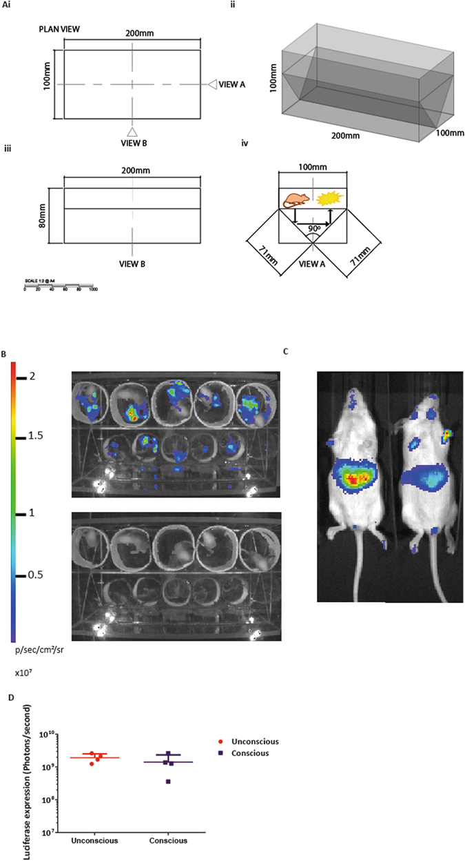
Whole-body bioluminescence imaging of conscious mice which have received a neonatal intravenous injection of VSV-G SFFV biosensor. (Ai) Plan view of the chamber, the scale bar represents millimetres (mm). (Aii) A 3D model with measurements (mm) of the Perspex box. (Aiii) A side-view B represents the length and depth of the chamber. (Aiv) Elevation view A shows the 90 degrees angle of the mirrors to construct a periscopic chamber to permit simultaneous collection of light emission from both ventral and dorsal surfaces, shown by the arrows. Mice were administered with VSV-G SFFV lentiviral vector intravenously at birth (n = 8). (B) Mice were imaged while conscious using the Perspex chamber, 5 at a time as neonates. The top image shows the whole-body bioluminescence and the lower image shows the mice in the chamber. The luciferase expression obtained from the conscious mice in the Perspex chamber was quantified by adding the expression profiles from the two planes. (C) Post weaning the mice were randomly split into two groups, where half of the mice were imaged unconsciously (not in the Perspex chamber). (D) There was no significant difference of luciferase expression between the conscious and unconscious mice, P = 0.657, mean +/− SD.