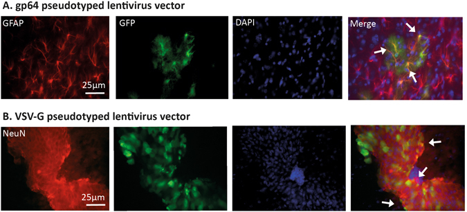
Figure 3.

Astrocytic targeting with gp64 pseudotyped lentivirus vector and neuronal targeting with VSV-G pseudotyped lentivirus vector. Brain sections from injected mice were used to determine the cell types expressing GFP after neonatal intracranial injections of gp64 or VSV-G SFFV lentivirus vector. (A) GFP with fibrotic GFAP astrocytic signals were co-localised. This is shown with the white arrows in the merge column. (B) GFP co-localised with NeuN in the CNS of mice which received the VSV-G SFFV vector (white arrows). All the images were taken at ×40 magnification.
