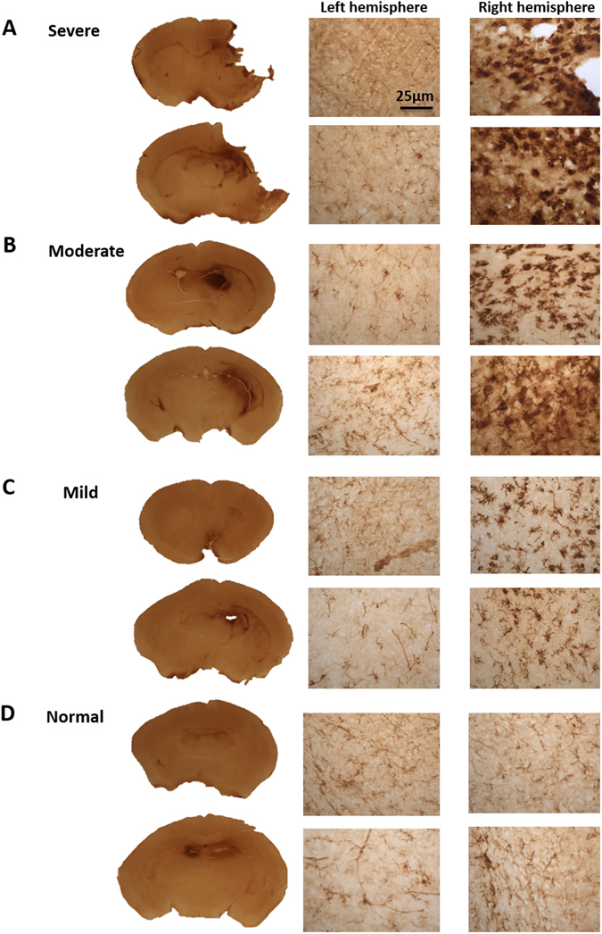
Figure 5.

Reactive microglia immunohistochemistry on HIE brains harvested 48 hours after surgery. Reactive microglia was detected by conducting a CD68 immunohistochemistry on the variable HIE brains. (A) Severe brains showed reactive microglia within the cortex and the hippocampus of the right lesioned hemisphere. CD68 positive cells were also found within the (B) moderate and (C) mild right hemisphere. Whereas CD68 expression was, absent from the (D) normal brain. The brain section images were taken at ×5 magnification and the left and right hemisphere images were taken at ×40 magnification.
