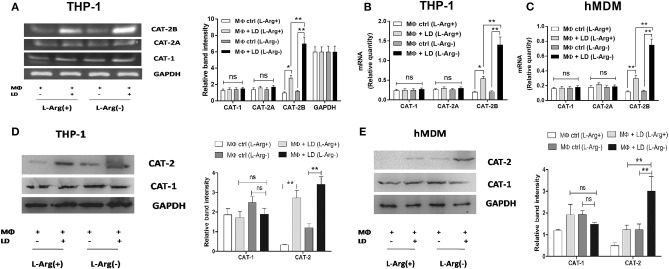
Figure 2.
Effect of Leishmania infection on expression of CAT-1 and CAT-2. THP-1 monocyte-derived MΦs (A,B,D) and human monocyte-derived macrophages (hMDM) (C,E) (1 × 10⁶ cells/ml) were grown in l-arginine-supplemented and l-arginine-depleted RPMI medium followed by infection with Leishmania donovani (multiplicity of infection 1:10) for 48 h. The expression of CAT-1 and CAT-2 (CAT-2A and CAT-2B) were evaluated at the transcript level by semi-quantitative RT-PCR (A) and real-time PCR (B,C) and protein level by western blotting (D,E). The intensity of the bands were quantified by densitometry in Quantity one software. Each experiment was repeated three times. Semi-QRT PCR gel images and western blots are representative of three experiments. Densitometric plots are shown adjacent to the gel images. Data are mean ± SD (n = 3) (*p < 0.05; **p < 0.001; ns, non-significant) (Mann–Whitney U test).

