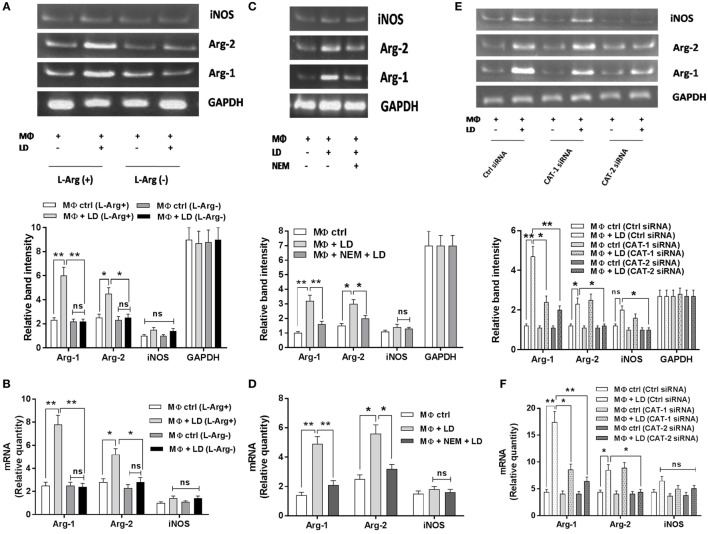
Figure 6.
Effect of inhibition of l-arginine availability on Arg-1, Arg-2, and inducible nitric oxide synthase (iNOS) expression in case of Leishmania infection. THP-1 monocyte-derived MΦs (1 × 10⁶ cells/ml) were grown in l-arginine-supplemented and l-arginine-depleted RPMI medium followed by infection with Leishmania donovani (multiplicity of infection 1:10) for 48 h (A,B). In another experiment, THP-1 monocyte-derived MΦs (1 × 10⁶ cells/ml) were left untreated or preincubated with NEM (250 µM) for 10 min followed by infection with L. donovani for 48 h (C,D). In a separate experiment, THP-1 monocyte-derived MΦs (1 × 10⁶ cells/ml) were transfected with control siRNA, CAT-1, or CAT-2 siRNA followed by infection with L. donovani for 48 h (E,F). The expression of Arg-1, Arg-2, and iNOS were determined at the transcript level by semi-quantitative RT-PCR (A,C,E) and real-time PCR (B,D,F). The intensity of the bands was quantified by densitometry using Quantity one software. Each experiment was repeated three times. Each determination was made in triplicate and the values were expressed as mean ± SD for three independent experiments. Semi-QRT PCR gel images are representative of three experiments. Densitometric plots are shown below the gel images. Kruskal–Wallis with Dunn’s multiple comparison test was used to evaluate statistical significance for comparing three or more groups; *p < 0.05; **p < 0.005; ns, non-significant.

