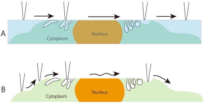
Figure 1.

An illustration showing the difference in imaging between resin sections (A) and cryosections (B) with AFM. A: Polymerized resin is hard to remove; therefore, the cantilever just traces the flat surface of the resin section. B: For the cryosection, the embedding medium (sucrose) was removed by immersing the sections in PBS. The fine structures in cells and tissues were then exposed, which enabled the cantilever to trace along any undulations that appeared.
