Figure 4.
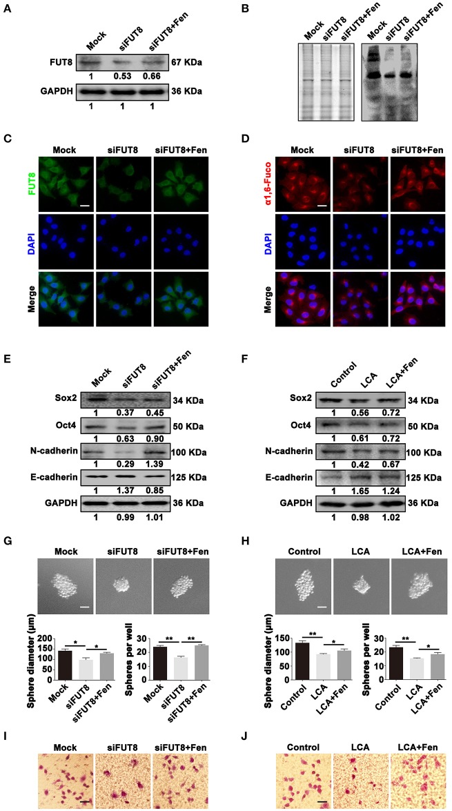
α1, 6-fucosylation is involved in fentanyl-mediated stemness and EMT. (A,B) Western blot, Lectin blot (C,D) immunofluorescent staining and Lectin fluorescent staining analysis for expression of FUT8 and α1, 6-fucosylation in mock, siFUT8 or siFUT8+fentanyl group. (E,F) Western blot analysis for Sox2, Oct4, N-cadherin and E-cadherin potein expression. (G) Representative images of tumor-spheres in mock, siFUT8 or siFUT8+fentanyl group observed under a microscope after 12 days of incubation. (Scale bars = 50 μm; Magnification, 200×). (H) Images of tumor-spheres in control, LCA or LCA+fentanyl group. (I) Images of migrated MCF-7 cells in mock, siFUT8 or siFUT8+fentanyl group observed under a microscope. (J) Images of migrated MCF-7 cells in control, LCA and LCA+fentanyl (0.1 μM) groups. (Scale bars = 50 μm; Magnification, 200×). Data are represented as mean ± SD (n = 3). Statistical significant different in sphere diameter and sphere per well (*P < 0.05; **P < 0.01).

