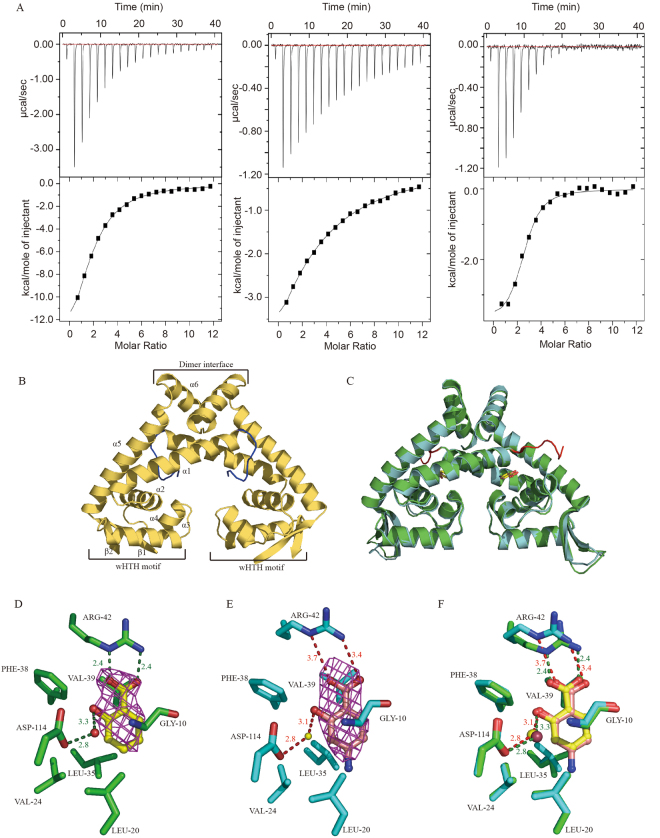
Figure 2.
Rv2887-SA and Rv2887-PAS complex structures show that SA and PAS bind to Rv2887 in a similar manner (A) Representative binding isotherms of SA (left) PAS (centre) and gemfibrozil (right) titrated into Rv2887, as determined by isothermal titration calorimetry. (B) Secondary structure of apo Rv2887. Elements of one of the subunits, the dimerization interface and the winged helix-turn-helix motif (wHTH) are labelled. (C) Secondary structure superposition of the Rv2887-PAS dimer (blue) and the Rv2887-SA dimer (green). (D) Stereo image of the SA binding pocket; residues from Rv2887 are shown as sticks. Polar contacts are shown as green dashed lines, and the red sphere is a water molecule. (E) Stereo image of the PAS binding pocket; residues from Rv2887 are shown as sticks. Ligands SA and PAS are surrounded by ligand-omit 1Fo-Fc electron density maps (magenta) contoured and 3.0 σ. Polar contacts are shown as red dashed lines, and the yellow sphere is a water molecule. (F) Stereo image of the superposition of the PAS binding pocket and the SA binding pocket.

