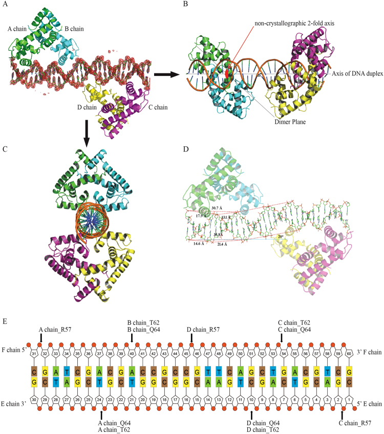
Figure 4.
The crystal structure of the Rv2887-DNA complex. (A,B,C) Orthographic views of the complex structure of a 30 bp DNA and two Rv2887 dimers in a unit cell. The DNA is surrounded by its ligand-omit 1FO-FC electron density map (red), contoured at 3.0 σ. The non-crystallographic 2-fold axis of the Rv2887 dimer is shown as a red ellipse and is perpendicular to the axis of the DNA duplex. The angle between the axis of the DNA duplex and the triangle-like shape of the protein dimer is ~40°. (D) The lengths of the major groove, the minor grove and the DNA duplex helix in one flank of the bound DNA are different from those in the other flank, and the angle of distortion is ~20°. (E) Schematic representation of the Rv2887-DNA interaction. Hydrogen bonds are indicated by arrows from the residues to the nucleotides.

