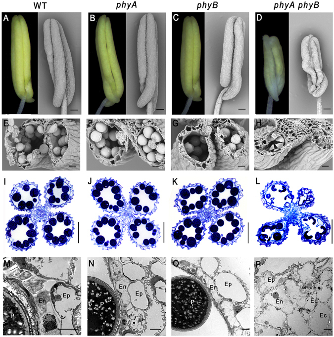
Figure 2.

Anthers of phyA and phyB single and double mutants, and wild-type (WT) rice at stage 12. (A–H) Light microscopic and scanning electron microscopic analysis of anthers at stage 12. (I–L) Sections of anthers at stage 12. (M–P) Transmission electron microscopic analysis of anthers at stage 12. Ep, epidermal cell layer; En, endothecium cell layer; Ec, extra cell layer; P, pollen grain. Bars = 0.1 mm in (A–D) 20 µm in (E–H) 50 µm in I–L, 5 µm in M–P.
