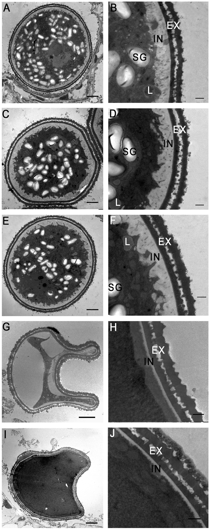
Figure 3.

Transmission electron microscopic analysis of pollen grains in the phyA and phyB single and double mutants, and wild-type (WT) rice at stage 12. Cross sections of pollen grains from WT (A and B), the phyA mutant (C and D), the phyB mutant (E and F), the phyA phyB mutant (G–J). EX, exine layer; IN, intine layer; SG, starch granule; L, lipid body. Bars = 5 µm in (A,C,E,G and I) 1 µm in (B,D,F,H and J).
