
Figure 3.
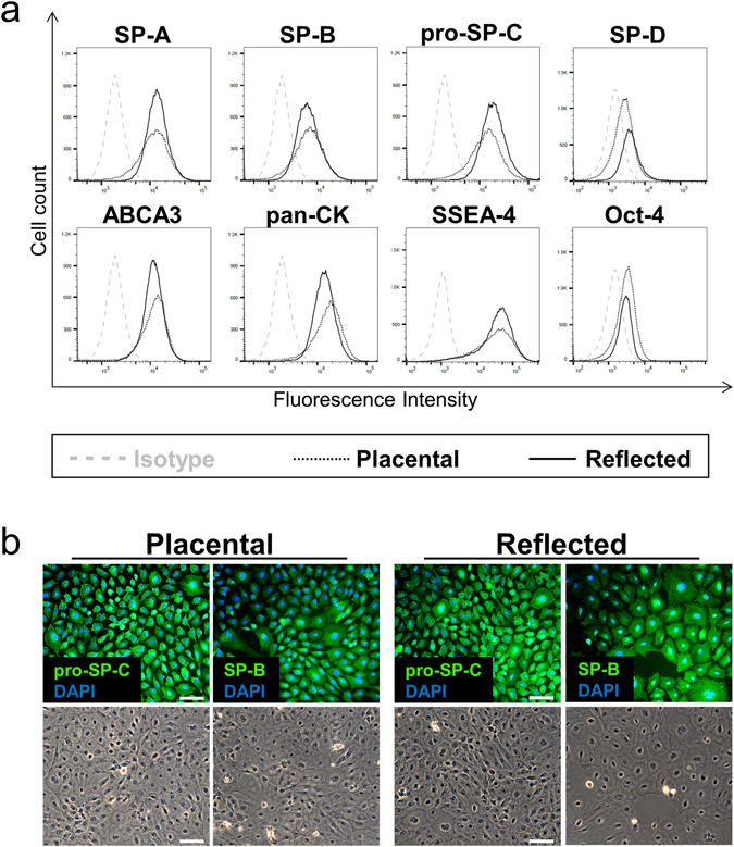
Expression of PS-associated proteins in isolated hAE cells. (a) Flow cytometric analysis of surfactant-associated proteins and stem cell markers immediately after hAE cells isolation. Cells show similar expression between both amniotic subregions, whereas in general hAE cells of the reflected region show moderately higher PS-associated protein expression and the placental area higher Oct-4 staining. n = 3–5. (b) Immunofluorescence staining of pro-SP-C and SP-B. The predominant part of hAM-derived epithelial cells exhibit distinct staining of both surfactant proteins in the cytoplasm, comparable in both amniotic subregions. Scale bars: 50 µm. n = 4.