Figure 3.
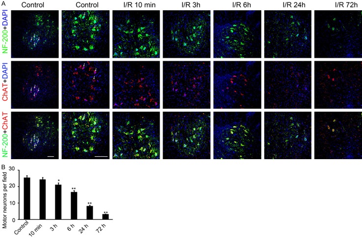
The expression of NF-200 and ChAT were detected in the ventral horn of spinal cord area on different time point post-operation. A. anti-NF-200 and anti-ChAT antibody were used for immunofluorescence in the ventral horn of spinal cord area. The green fluorescence signal indicates NF-200 protein and red fluorescence signal indicates ChAT protein. The nuclear was stained by DAPI. The tissues form control group, and 10 min, 3 h, 6 h, 24 h and 72 h post operation group was collected and used for staining. The double positive cells were labeled with white arrow. Scale bar: 150 μm. B. The double positive cells in per field were counted and analyzed. (n=40, *P<0.05, **P<0.01, compared with control group).

