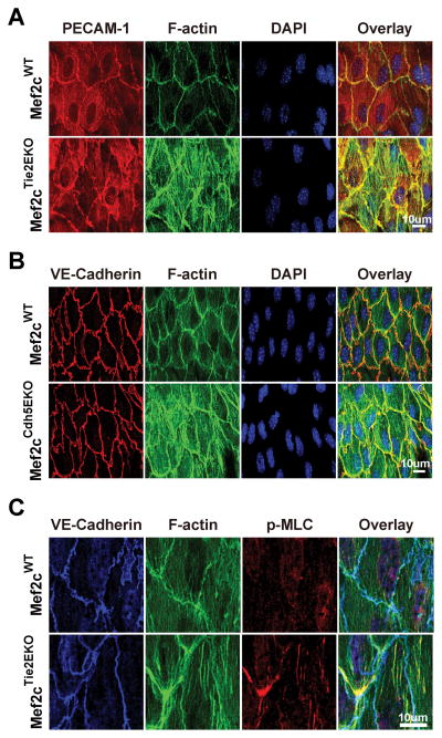
Figure 2. Endothelial Mef2c deletion leads to MLC phosphorylation and actin stress-fiber formation in the thoracic aorta.
En face preparation and immunostaining were performed on the thoracic aorta as described in methods. A, B, thoracic aortas from Mef2cTie2EKO and control Mef2cWT mice were immunostained for PECAM-1, F-actin (phalloidin), and nuclei (DAPI). B, Mef2cCdh5EKO (14-days post tamoxifen) and vehicle control were immunostained for VE-Cadherin, F-actin (phalloidin) and nuclei (DAPI); C, thoracic aortas from Mef2cTie2EKO and Mef2cWT control were immunostained for VE-Cadherin, p-MLC, and F-actin (phalloidin). Representative confocal images are shown (n=4).

