
Figure 3.
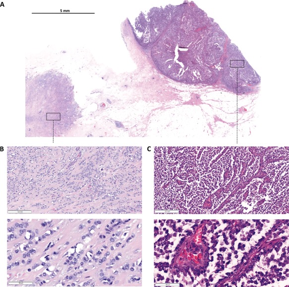
Resection specimen. (A) Submacroscopic view of a representative HE stained FFPE tissue block covering the margin of the main tumour and its capsule (right, solid‐papillary‐like growth pattern) and an adjacent satellite focus (left, classical lobular growth pattern). (B and C) Details at x50 and x200 magnification.