Figure 4.

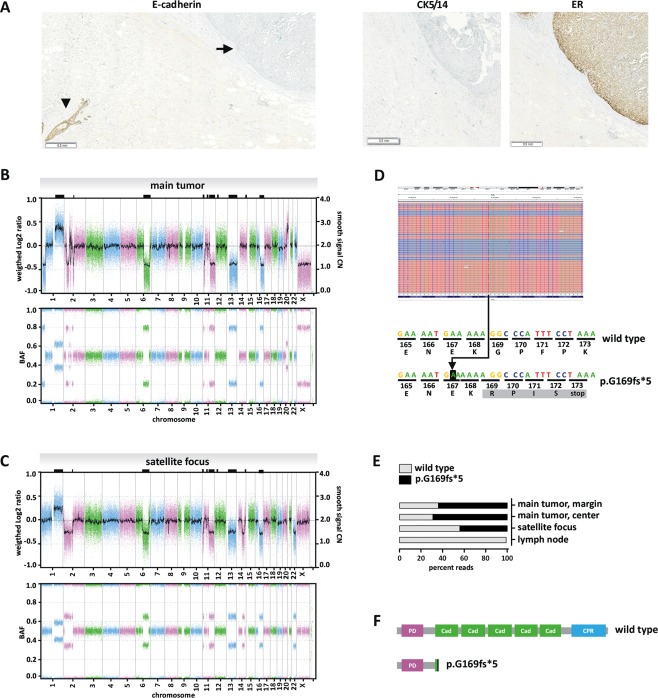
Molecular characteristics. (A) Loss of E‐cadherin protein expression in the solid‐papillary‐like main tumour. The FFPE block shown in Figure 2 was subjected to E‐cadherin immunohistochemical staining. E‐cadherin immunoreactivity was completely lost in the main tumour (closed arrow), while an adjacent normal mammary gland duct was strongly positive for E‐cadherin, which served as internal positive control (arrow head). Please note that the slide is presented at a submacroscopic view to demonstrate that the loss of E‐cadherin expression was complete and uniform. Immunohistochemical staining for ER is provided for comparison (right). Staining for CK5/14 is provided to demonstrate that the main tumour lacked a myoepithelial cell layer along the capsule. (B) Array‐based DNA copy number profiling of the main tumour with solid‐papillary‐like growth pattern. The upper panel presents a whole‐genome overview of copy number alterations (CNAs) with chromosomal locations plotted on the x‐axis. The weighted Log2 ratios and copy numbers (represented as a Gaussian smoothed calibrated copy number estimate) are plotted on the left and right y‐axis, respectively. The lower panel presents the corresponding B‐allele frequencies (BAF). (C) array‐based DNA copy number profiling of the satellite focus with classical lobular growth pattern. Shared CNAs are highlighted by black rectangles on the upper frame of the plot. (D) CDH1 mutational analysis by NGS. Sequencing reads of DNA from the solid‐papillary‐like main tumour were aligned with the IGV Browser, version 2.3.78 (upper panel). Reverse reads are shaded in blue, forward reads in red. Reads with an insertion variant are highlighted by a blue mark (upper panel). Sequence details are shown in the lower panel. The 'A' on black background corresponds to a c.499_500insA insertion mutation. The resulting frameshift generates an altered protein sequence (grey background) and a premature stop at codon 173. (E) Percentage of NGS reads carrying the c.499_500insA mutation in different DNA preparations. (F) Schematic presentation of the human E‐cadherin protein. PD; prodomain, Cad; cadherin domain; CPR, cytoplasmic region. The c.499_500insA mutation results in a heavily truncated E‐cadherin protein (p.G169fs*5).
