
Figure 3.
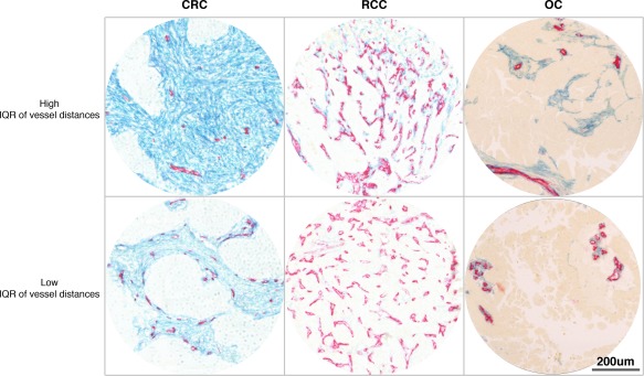
CRC, RCC, and OC cases with high or low inter‐vessel distance heterogeneity (vessel distance IQR). Photomicrographs showing examples of CRC (left), RCC (middle), and OC (right) tumours with high (upper) or low (lower) heterogeneity regarding inter‐vessel distances. Sections have been stained to detect endothelial cells with CD34 antibodies (red) and with PDGFR‐β antibodies (blue).