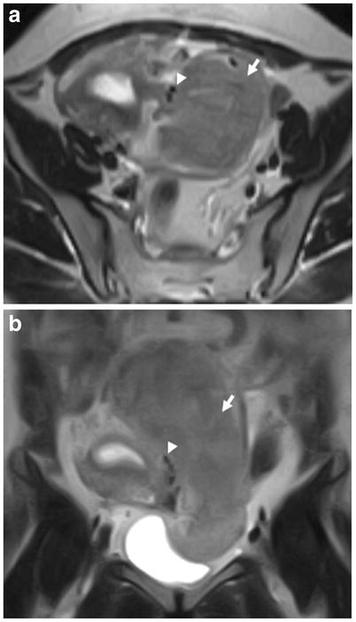
Figure 3.

Axial T2-weighted (a) and coronal T2-weighted (b) images from a MRI (low-field-strength magnet) obtained outside our institution in a 38-year-old woman with a pelvic mass seen on ultrasound. The initial MRI report described a large left pelvic mass (arrow) suspicious for an adnexal neoplastic process of indeterminate malignant potential. The second-opinion MRI interpretation by a gynaecologic oncologic radiologist characterized this mass as a subserosal leiomyoma based on the presence of multiple vessels (arrowhead) between the uterus and a juxta-uterine mass (i.e. bridging vascular sign). The diagnosis of a subserosal leiomyoma was confirmed at the subsequent surgery. Second-opinion review was correct despite the limited quality of the study related to the low-field-strength of the magnet.
