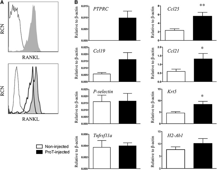
Figure 6.
Gene expression analyses from thymuses obtained from in vitro–derived proT-injected mice. (A) Flow cytometric analysis of RANK ligand (RANKL) expression on purified CD34+CD38− cells (not shaded) and day 11 in vitro–derived CD34+CD7+-gated proT-cells (shaded) (top); and in vitro–derived proT1- (not shaded, thick line) and proT2-cells (shaded) (bottom). Unstained cells are included as a control (not shaded, thin dashed line). (B) Quantitative real time reverse transcriptase polymerase chain reaction analysis for the expression of human PTPRC (CD45), and mouse Ccl25, Ccl19, Ccl21, Selp (P-selectin), Krt5 (Cytokeratin-5), Tnfrsf11a (RANK), and H2-Ab1 (MHC class II) from mouse thymus extracts of NSG mice injected with proT-cells or control noninjected mice after 3 weeks. Transcript levels for all genes were normalized to mouse β-actin. These results are the average of 3 independent experiments, with the exception of Selp (n = 2), with error bars corresponding to standard error of the mean. Asterisks represent statistical significance as determined by Student t tests. The results shown are representative of 3 independent experiments. *P < .05; **P < .005.

