FIG 1.
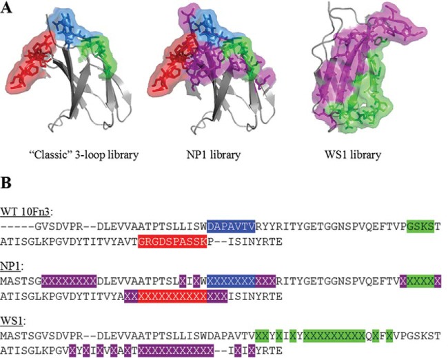
Adnectin library designs. (A) Depiction of randomized regions of the 10Fn3 domain within the classic (left), NP1 (center), and WS1 (right) libraries. The classic and NP1 libraries have the BC loop in blue, the DE loop in green, and the FG loop in red. Flanking and N-terminal randomized residues in the NP1 library are shown in purple. The WS1 structure is turned so that the B-C-F-G β-sheet is facing front. In the WS1 structure, randomized residues in the CD strands and loop are green, while those in the FG strands and loop are purple. The structure was taken from the protein with PDB accession number 1FNF. (B) Comparison of the sequences of the WS1 and NP1 libraries with the sequence of the wild-type (WT) 10Fn3 domain. Highlighted sequences in wild-type 10Fn3 indicate the residues that are randomized in the classic library. The color coding is as described in the legend to panel A. Highlighted X's indicate randomized residues in the new libraries. Dashes indicate sequence gaps to facilitate visual alignment across the libraries.

