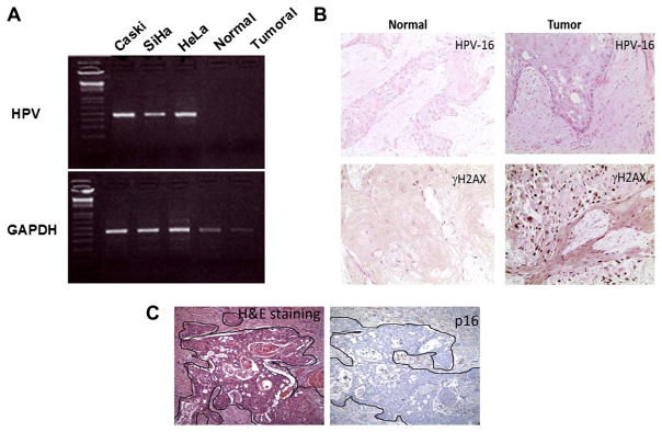
Fig. 2.
Absence of HPV detection in tumour biopsy. (A) PCR amplification of the genomic DNA for HPV 16 and HPV 18 shows that both normal and tumoural tissues from the patient are negative for HPV, while cell lines with integrated HPV 16 or HPV 18 show the correct size band for HPV. (B) In situ hybridisation confirms the HPV negative status of the patient. γH2AX, a marker for genomic instability, was used as a positive control. (C) A staining for p16 confirms the absence of the protein in both normal and tumour tissues.

