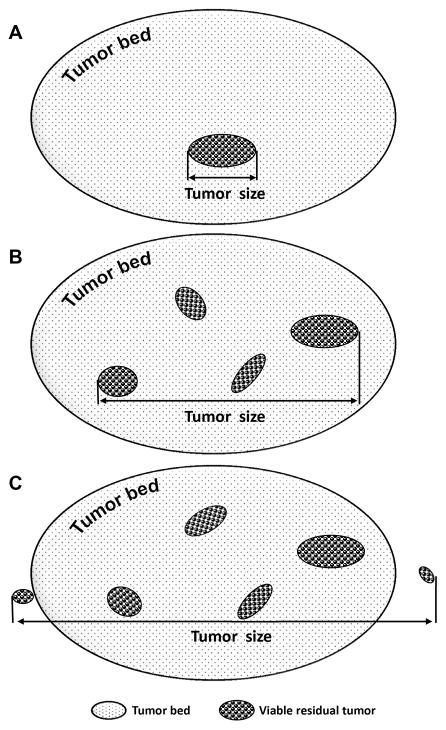
Figure 1.
Schematic drawing to illustrate the method for tumor size measurement. For cases which had only single microscopic focus of viable residual tumor, the largest linear dimension of the viable tumor focus on H & E slide was used as the final tumor size (A). For cases which had more than one microscopic foci (multifocal) of viable residual tumor, the largest linear dimension of the area involved by all islands of viable residual tumor and the intervening fibrotic stroma was used as the residual tumor size (B). For cases which had microscopic viable residual tumor invading the pancreas and/or soft tissue beyond the tumor bed area, the largest dimension of the entire area involved by all islands of viable residual tumor with the intervening stroma, pancreas and/or soft tissue was used as the final tumor size (C).

