
Figure 2.
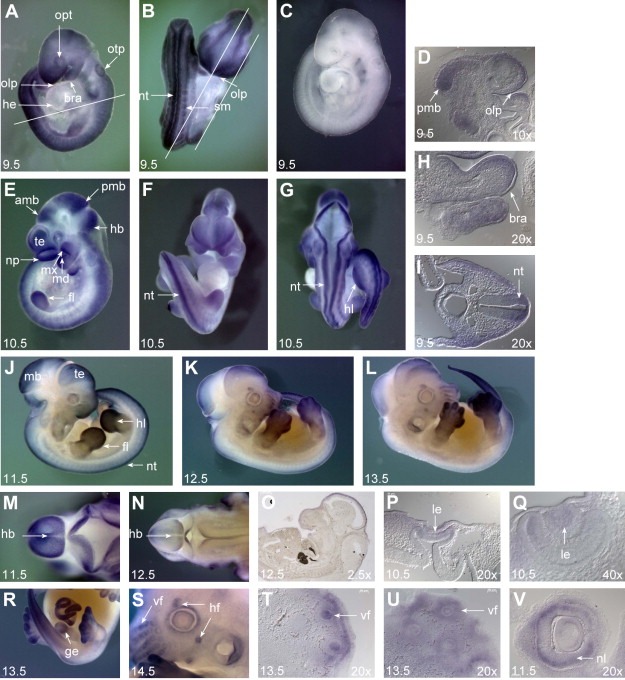
Whole mount in situ hybridization analysis during mouse embryonic development. Embryos were hybridized with the Cats antisense probe. (A, B, D, H, I) Expression of Cats at embryonic stage of 9.5d.p.c. Widespread transcript distribution with prominent expression in the neural tube (nt), somites (sm), posterior region of the midbrain (pmb), olfactory placode (olp) and in branchial arches (bra). Note the complete absence of Cats expression in the primitive heart (he). Otic pit: otp; optic vesicle: opt. Bars indicate the plane of section. (C) 9.5d.p.c. embryo hybridized with the Cats sense probe as a control for staining specificity. (E–G, P, Q) At 10.5d.p.c., there is a reduction of widespread Cats expression, transcripts are additionally detected in fore‐ and hindlimbs (fl and hl, respectively), telencephalon (te), nasal process (np), lens vesicle (le), anterior region of the midbrain (amb), hindbrain (hb) and mandibular and maxillary component of first branchial arch (md and mx, respectively). (J–L) Right lateral view of embryos from 11.5, 12.5 and 13.5d.p.c. Note reduction of Cats overall expression and prominent expression in the limbs. (M, N) Dorsal view of the cephalic region of 11.5 and 12.5d.p.c. embryos. Note the abrupt reduction of Cats expression in the hindbrain and neural tube from stage 11.5 to 12.5d.p.c. (O) Remaining Cats expression in the midbrain of a 12.5 embryo. (P–V) Transcripts detected in the lense vesicle (le), genital tubercle (ge), hair follicles (hf), vibrissae follicles (vf) and neural layer of optic cup (nl). (D, H, I, O–Q, T–V) Sagittal (D, H, O, T–V) and transverse (I, P, Q) sections from whole mount stained embryos. Stage of development and imaging magnification are indicated on the left and right bottom of the photographs, respectively.