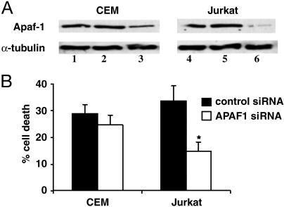
Fig. 7.
Knockdown of APAF1 expression attenuates SAHA-induced apoptosis. (A) CEM and Jurkat cells transfected with no siRNA (lanes 1 and 4), 1.25 μM control (GFP) (lanes 2 and 5), or APAF1 siRNA (lanes 3 and 6) were analyzed 48 h after transfection for APAF1 expression by Western blot. The blot was reprobed for α-tubulin as a loading control. (B) At 48 h after electroporation, CEM and Jurkat cells were treated with SAHA (2.5 μM) for 18 h, then collected and scored for apoptosis by propidium iodide staining. Data are expressed as the mean ± SD of three separate experiments. Statistical differences between samples (P < 0.05) were determined by using the Mann-Whitney U test and are denoted by an asterisk.

