
Figure 3.
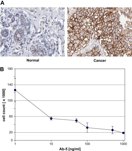
Immunohistochemistry and cell growth inhibition. Ab–5 was analyzed by immunohistochemistry on normal and cancer tissues (Agaton et al., 2004; Nilsson et al., 2005). Weak membranous/cytoplasmic staining can be seen on normal breast glandular cells, in contrast to a clear membranous staining for breast ductal carcinoma over‐expressing HER2 (A). A dose‐dependent effect on cell growth of breast cancer cells treated with increasing concentration of antibody Ab–5 was shown in a 5day titration study using human BT474 cells (B).