
Figure 6.
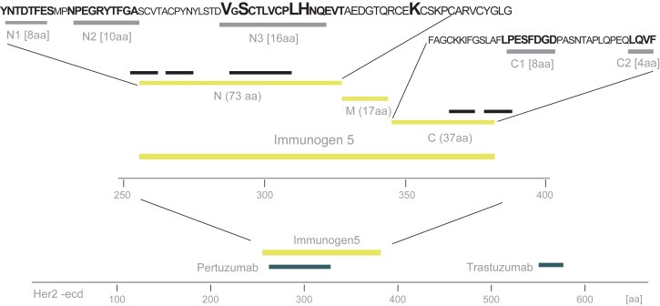
In‐depth epitope mapping of antibody sub‐fractions. Ab–N and Ab–C were mapped using a bacterial cell display system (Rockberg et al., 2008). The extracellular domain is shown with the approximate binding regions of the two monoclonal antibodies pertuzumab and trastuzumab. Immunogen 5 and its three sub regions (N, M and C) cover a stretch of 127 aa and the epitope mapping revealed consensus sequence depicted as N1, N2, N3 on the N‐terminal region and C1 and C2 on the C‐terminal region (see also Supplementary Figure 2). Four of the residues in epitope N3 (emphasized) showed to be residues in the previously published (Franklin et al., 2004) pertuzumab epitope.