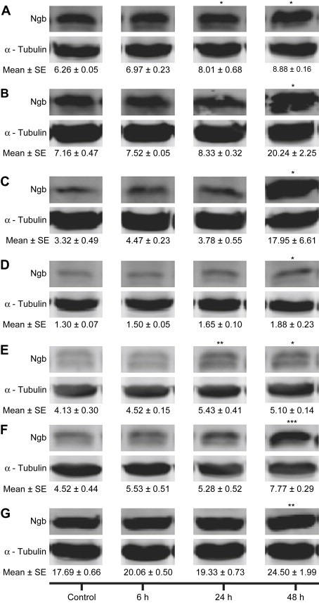
Figure 2.

Ngb protein expression in human GBM cells. (A) M010B; (B) M059J; (C) M006x; (D) M006xLo; (E) M059K; (F) U87R; (G) U87T. Ngb expression was assessed by Western blot analyses after exposure to hypoxia (0.6% O2) for 0, 6, 24 and 48h. The integrated intensities of Ngb and α‐tubulin (control) bands were determined and expressed in arbitrary units (AU), and representative blots are shown. (n=4) *P<0.05; **P<0.01; ***P<0.001 (ANOVA).
